
Overview
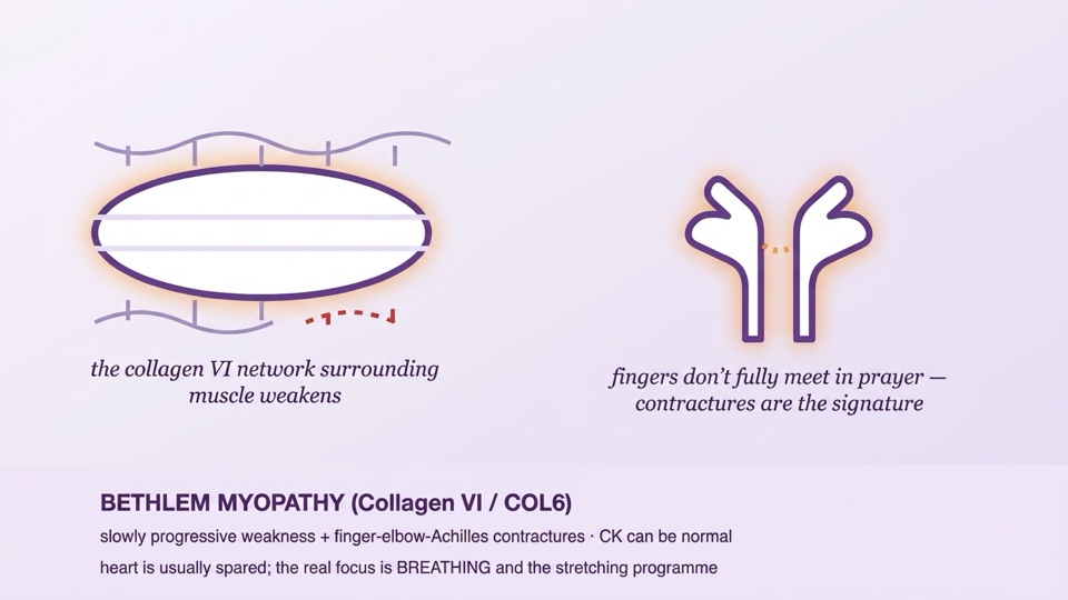
Bethlem myopathy is a slowly progressive muscle disease caused by weakening of the connective tissue network surrounding muscle (collagen VI; the COL6A1, COL6A2, COL6A3 genes). Its signature is not so much weakness as contractures: shortening of the long finger flexors ("the palms do not fully meet when praying"), and stiffness at the elbows and Achilles tendon. The severe end of the same genes is Ullrich congenital muscular dystrophy (early severe weakness combined with the striking coexistence of joint laxity and stiffness); Bethlem is the end of this spectrum in which children walk, go to school, and often remain active even into adulthood. It carries two critical messages: a normal CK does not rule out muscle disease, and while the heart is generally spared, it is the respiratory system that truly needs monitoring — the diaphragm can be silently affected even while the patient is still walking.
Genetic Cause and Mechanism
- Collagen VI is a microfibrillar network that anchors the muscle fibre to the extracellular matrix; when disrupted, the muscle-matrix connection and the muscle's repair signalling are weakened.
- Inheritance is mostly autosomal dominant (familial or de novo; dominant-negative variants are common); recessive forms also occur — parental examination (finger contractures, skin findings) is part of family counselling: a mildly affected parent is easily overlooked, and the transmission risk is 50%.
- If the genetic report shows a VUS, reclassification should be sought every 1-2 years; in a classic clinical picture with a negative panel, further investigation (RNA studies) for deep-intronic variants may be considered.
Clinical Picture
- Onset: Often subtle in infancy-childhood: hypotonia, torticollis, or transient joint laxity in infancy; in some children only "clumsiness" and difficulty with stairs.
- Contractures — the signature finding: long finger flexors (fingers do not reach full extension when the hands are raised), elbows and Achilles; a description of stiffness lasting through the night is typical. Contractures may precede weakness.
- Weakness: proximal, mild-to-moderate, and slowly progressive; a proportion of adults need walking support at an advanced age.
- Skin clues: keloid/atrophic scars and follicular hyperkeratosis ("keratosis pilaris" / "goose skin") — these are not mentioned unless asked about on examination, and are a shortcut to diagnosis.
- Respiratory system: late and mild in Bethlem, but diaphragm involvement can develop even while walking is preserved (early and prominent at the Ullrich end): morning headache, daytime sleepiness, and frequent night-time awakening are questions pointing to nocturnal hypoventilation.
- Heart: typically spared — an important difference from Duchenne; beyond a baseline assessment, routine progressive cardiac follow-up is not required (if there are no findings).
- CK: normal or mildly elevated (often ≤5-fold) — one should not fall into the trap of "CK is normal, therefore it is not a myopathy".
Diagnosis
- The clinical pattern (contracture + slow weakness + skin findings) raises suspicion; the muscle MRI/ultrasound pattern supports it (described appearances such as outside-in involvement in the thigh muscles and a "central shadow" in the rectus femoris).
- Definitive diagnosis is genetic: COL6A1-3 panel/exome; if necessary, collagen VI studies on skin fibroblasts are supportive.
- Baseline package: a measured map of joint ranges, pulmonary function (at an age the child can perform it — sitting and supine FVC; a supine drop is a clue to diaphragm involvement), a one-time baseline ECG-echocardiogram, and skin assessment.
Treatment — An Honest Framework
- The core of treatment is a physiotherapy-stretching programme: regular stretching plus night-time orthoses slow the progression of contractures; surgical lengthening may be considered in selected cases of severe Achilles contracture (followed by early mobilisation).
- Respiratory system: annual follow-up; a sleep assessment when there are findings/suspicion of nocturnal hypoventilation, and night-time NIV when needed — this notably improves quality of life.
- Anaesthesia note: in the context of neuromuscular disease, avoiding succinylcholine and careful planning is the safe approach; the diagnosis must always be disclosed before a procedure.
- Exercise: regular low-to-moderate intensity activity that avoids eccentric loading is recommended; prolonged immobilisation accelerates contractures — early movement should be planned after surgery/casting.
- Research horizon: allele-specific ASO approaches aimed at silencing dominant-negative COL6 variants are at the preclinical stage; all of these are at the research stage and are not a recommendation for clinical use. Collagen VI patient registries keep the door open to studies.
Three Critical Points in the Emergency Department
- 1 · Respiratory system: have a low threshold for hypoventilation in lower respiratory tract infection; in advanced cases avoid giving oxygen alone without blood gas monitoring (prioritise NIV).
- 2 · Anaesthesia: succinylcholine is not used; a postoperative respiratory and early mobilisation plan is needed (immobilisation accelerates contractures).
- 3 · CK may be mildly elevated — this should not be mistaken on its own for acute muscle breakdown; cardiac involvement is not expected, and cardiac alarm is unnecessary if there are no symptoms.
Follow-up — Summary
The backbone of follow-up: measuring the joint-contracture map every 6-12 months plus updating the stretching programme and night-time orthoses · annual respiratory follow-up (sitting + supine FVC; sleep assessment if symptomatic) · scoliosis examination · a one-time baseline ECG-echo (repeated if new symptoms arise) · skin (keloid tendency — noted for planned surgeries) · weight-nutrition · adaptation of physical education at school · family examination (mildly affected parent) plus genetic counselling and VUS reclassification. The detailed year-by-year table is in the Routine Follow-up Calendar document at the end of this article.
Prognosis — An Honest Framework for Families
- Bethlem is at the slow and relatively benign end of muscle diseases: childhood and young adulthood are mostly spent with independent walking; a proportion may need a cane/walking aid at an advanced age.
- Life expectancy is generally normal; the main concerns of the later period are respiratory and contracture management — both of these are predictable with follow-up.
- The picture is earlier and more severe at the Ullrich end (loss of walking, early night-time NIV); both ends of the spectrum can occur together in the same family.
Daily Life
- Build the stretching routine into daily life (10 minutes after bathing); use night-time orthoses regularly.
- Add the questions "morning headache / daytime drowsiness" to the annual checklist — these are early signals of nocturnal breathing problems.
- Disclose the diagnosis and the succinylcholine note before every surgery-anaesthesia; request early physiotherapy after casting/rest.
- Share the keloid tendency of scars with your surgeons.
- Join collagen VI family communities and registries.
Downloadable Documents: Emergency Action Plan + Routine Follow-up Calendar (PDF)
Two documents have been prepared by Nörogender, under the guidance of Prof. Dr. Burak Tatlı, for children diagnosed with Bethlem/collagen VI disorders. The Emergency Action Plan (3 pages): alongside sick-day steps and thresholds for seeking care, it includes notes on avoiding succinylcholine, a warning against unmonitored oxygen in advanced cases, "CK may be mildly elevated", and early mobilisation. The Routine Follow-up Calendar (2 pages): a year-by-year table and record chart built around the contracture map, sitting+supine FVC, questioning for nocturnal hypoventilation, and the stretching-orthosis programme.
📄 Emergency Action Plan (PDF, 3 pages) — in Turkish 📅 Routine Follow-up Calendar (PDF, 2 pages) — in Turkish
Complete the documents together with your physician; keep a copy at home and at school.