
Overview
One of the sentences families hear most often from the genetics laboratory is: "The whole genome was sequenced, nothing came up." This sentence is often understood as "so my child's condition is not genetic". Yet a negative genome report is not a biological result; it is the record of an investigation carried out on a particular sample, with a particular technology, with a particular bioinformatics pipeline, and with the knowledge available on the date the report was written. If clinical suspicion continues, the evaluation is not over.
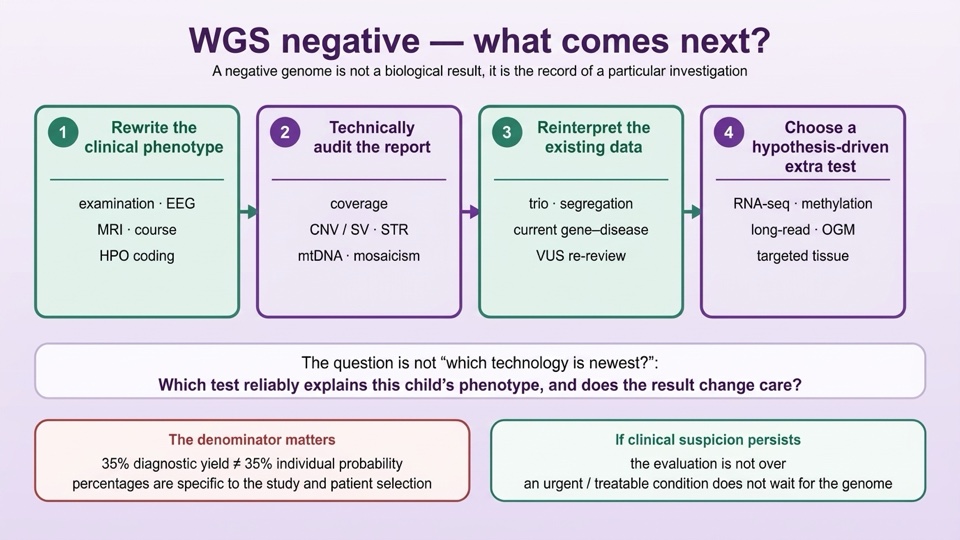
This article was written for children with strong suspicion of an inherited disease in whom exome (WES) or genome (WGS) sequencing has not led to a diagnosis: children with epilepsy, developmental epileptic encephalopathy, global developmental delay, intellectual disability, syndromic autism, movement disorder, ataxia, spasticity, hypotonia and cerebral palsy phenotype. The aim is not to list new tests; it is to audit the limits of the first investigation, rewrite the phenotype, and choose only the method that fits the clinical hypothesis.
What a "negative" result means, and what it does not
For a molecular diagnosis, a pathogenic or likely pathogenic variant must be consistent with the phenotype and the inheritance pattern. A variant of uncertain clinical significance (VUS) may be a strong candidate, but on its own it is not considered a definitive diagnosis. A negative report, on the other hand, is an exclusion limited only to the variant classes the test is sensitive to. Demonstrating a change in the genome is also not in itself proof of causality: segregation, RNA effect, functional evidence and support from the literature are sought.
Clinical decisions are not hostage to this report. If progressive neurological deterioration, acute encephalopathy, or a treatable metabolic condition is considered, parallel urgent diagnostic and therapeutic procedures are carried out without waiting for the genomic result.
WES and WGS are not the same test
WES mostly targets exons and adjacent intronic regions; it examines approximately 1–2% of the genome at high read depth. WGS reads a much larger part of the genome and, with suitable pipelines, is suitable for evaluating intronic regulatory variants, copy number variants (CNVs), structural variants (SVs) and repeat expansions. Even so, standard short-read WGS cannot fully resolve every locus.
| Feature | WES | Short-read WGS |
|---|---|---|
| Primary content | Exon + nearby splice | Broad coding and non-coding genome |
| Depth | Higher at target region | Lower but homogeneous |
| SNV / small indel | Strong | Strong |
| SV / CNV | Analysis-dependent, limited | Better; caller-dependent |
| Repeat expansion (STR) | Generally limited | Can be partly resolved with suitable algorithm |
| mtDNA / low-level mosaicism | Depends on targeted conditions | Standard analysis may not be sufficient |
Mother–father–child (trio) sampling strengthens the interpretation of de novo variants, trans position in recessive disease, and X-linked inheritance. In a patient whose phenotype has been very well characterised, a single-sample WGS may give a more meaningful result than expected.
When reading percentages, ask about the denominator
A large meta-analysis dated 2025 covered 416 studies and 124,937 participants. For neurodevelopmental disorders, it reported pooled diagnostic yields of 14.8% for chromosomal microarray, 35.3% for WES, and 30.5% for WGS. In epilepsy these figures were 13.3%, 34.2% and 39.1% respectively. These percentages are not head-to-head comparisons made in the same children; they come from different centres and different patient selections, and heterogeneity between studies is high. Indeed, in a 2025 paediatric rare disease meta-analysis, within the same cohort, genome sequencing was found to yield 30.6% and exome sequencing 23.2%; there was a trend favouring genome, but the difference was not statistically definitive.
More importantly: a 35% diagnostic yield does not mean "this child's disease has a 35% probability of being genetic". This figure reflects the known molecular causes the test captures, patient selection, and the laboratory's analytical scope. When reading a percentage, ask three things together: whether it is the initial yield or the additional yield in a subgroup whose previous tests were negative, in which patient type, and with which test definition.
Why does a genome remain "negative"?
The reasons fall under two headings. Technical and bioinformatic limits: low read depth, GC-rich sequences and alignment errors can mask some loci; standard short reads may fail to resolve large balanced translocations, inversions and complex structural variant breakpoints; a suitable specialised caller may not have been used for repeat expansions; CNV, uniparental disomy, mtDNA and heteroplasmy workflows may not have been within scope from the outset; a blood sample may not represent somatic mosaicism limited to a particular tissue.
Interpretation and biology limits: the effect of a deep intronic variant may not be demonstrable from DNA data alone; the gene–disease relationship may have been defined after the date of the report; a candidate variant may have been filtered out as a VUS and never reported to the clinician; if phenotype data is incompletely coded, the correct variant may rank lower in prioritisation; the genetic model may not be monogenic — imprinting, methylation, digenic effect, or polygenic susceptibility may be involved.
In practice this distinction becomes the question: is this negativity technical, or biological? The PDF report alone is not enough to answer this; the test's methods documentation, caller list, coverage metrics and, where necessary, raw data files are evaluated. The most common practical mistake is to request a new and costly test straight after a negative report; the relevant structural variant, repeat, or mitochondrial analysis may never have been performed in the first study at all.
The cheapest and most powerful step: rewriting the phenotype
A negative genome benefits most from new clinical observation and reinterpretation. The phenotype changes as the child grows: a child initially recorded as "hypotonia + developmental delay" may show dystonia at 2 years of age, ataxia at 4 years, and peripheral neuropathy may become apparent later in the course. EEG evolution, seizure type, the distinction between leukomalacia and malformation on MRI pattern, and whether the course is progressive or not, reshape the clinical hypothesis.
For this reason, it is useful to produce a one-page phenotype summary at the second evaluation: pregnancy and birth, family history, skills gained and lost, motor phenotype, seizure phenomenology, cognition–language, behaviour, head circumference, sensory systems, imaging, disease course and previous molecular results. Rather than the label "global developmental delay" copied from the report, writing explicitly, for example, that apraxia is prominent while social communication is preserved, or that white matter signal is regressing, can contribute directly to the diagnosis.
Reanalysis or a new test?
These two options are not opposites; their order is determined according to the patient. In the 2025 meta-analysis, reanalysis of existing WES/WGS data provided an additional 10.6% diagnoses in the neurodevelopmental disorder cohort and an additional 10.4% in the epilepsy cohort. In children with rare disease whose previous WES was negative, reanalysis of WES gave 14.2% additional diagnoses, while new findings specific to WGS gave 7.0%. These different percentages represent different methods, denominators and case selections; there is no single universal additional-yield percentage after a negative WGS.
The practical decision is built as follows: if the relevant gene was well covered and there is a strong intronic VUS, RNA testing may be more meaningful than repeating WGS. If the previous WES was interpreted with an older panel and older callers, reanalysis of the raw data, CNV calling and the advantage of trio sequencing come to the fore. If the hypothesis of a structural variant, repeat expansion, or non-coding region is strong and the previous test did not cover this, the move is to genome-level or targeted advanced methods.
Which method for which suspicion
Tests are not a mandatory sequence; different mechanisms can be investigated in parallel in the same patient.
| Clinical hypothesis | Preferred method | What it shows |
|---|---|---|
| Intronic / exon-edge VUS predicted to affect splicing | RNA sequencing (RNA-seq) | Exon skipping, cryptic splicing, expression level |
| Chromatinopathy / syndromic developmental delay | DNA methylation signature (episignature) | Syndrome-specific epigenetic pattern |
| Structural variant, repeat expansion, complex locus | Long-read WGS or optical genome mapping | Breakpoint, phase, repeat size |
| Somatic mosaicism or mitochondrial disease | Deep targeted analysis in the appropriate tissue | Low allele fraction, mtDNA heteroplasmy |
| Treatable metabolic condition | Targeted biochemistry and metabolomics | Functional evidence of a disrupted pathway |
Each of these methods carries its own limits. RNA testing is only meaningful in the tissue that expresses that gene; blood, fibroblast and muscle profiles cannot substitute for one another. A methylation signature is only exclusionary for syndromes whose signature has been defined. Long-read and optical mapping provide additional diagnoses in selected unresolved cohorts; no study recommends them as "routine second-line testing in all WGS-negative children". A negative blood WGS does not exclude a tissue-limited somatic variant or a particular mtDNA heteroplasmy.
A special situation in cerebral palsy and autism
Acquired injury does not in itself exclude a genetic contribution. In a JAMA Pediatrics meta-analysis dated September 2026, overall genetic testing yield was 19% in unselected cerebral palsy groups; 44% in cryptogenic cerebral palsy with unexplained aetiology; and 13% in non-cryptogenic cases. Red flags that prioritise evaluation are: normal or non-specific brain MRI, or an MRI lesion disproportionate to the clinical motor findings; progressive weakness, loss of acquired skills, predominant ataxia or dystonia; dysmorphic features, congenital anomalies, similar disease in the family; a weak perinatal explanation.
The co-occurrence of cerebral palsy and autism is also a point frequently confused. In a 2024 US population-based analysis, autism prevalence was 6.09% in children with a cerebral palsy diagnosis, versus 1.15% in those without. This shows that the clinical co-occurrence of the two conditions is increased; it is not the detection rate of two separate molecular diseases. Truly having two separate Mendelian diseases in the same child ("dual molecular diagnosis") is much rarer: it has been reported in 4.2% of those diagnosed in a cohort selected from severe and unresolved rare diseases. Reasonable grounds for seeking a second diagnosis are: the first molecular diagnosis explaining only part of the clinical findings, finding organ involvement that is biologically inconsistent with the existing diagnosis, or seeing a split clinical pattern in the family.
Questions for families and physicians to ask
Before requesting a new test, auditing the existing report is often more productive. The following questions can be answered together with the laboratory and the clinician:
- Was trio sequencing performed; were family history and consanguinity included in the model?
- Was coverage adequate at risk loci; which regions could not be read?
- Were copy number variants, inversions and translocations looked for?
- Is there a specialised caller or confirmation for a repeat expansion (for example FMR1) appropriate to the phenotype?
- Were mtDNA heteroplasmy and nuclear mitochondrial genes evaluated?
- Was sensitivity for low allele fraction mosaicism reported; was the appropriate tissue chosen?
- Have candidate variants that were consistent with the phenotype but not reported been re-examined?
- Have current neurological, ophthalmological, audiological and imaging findings been added to the analysis?
- What is the policy on raw data access, storage duration, and reinterpretation?
The value of this audit does not come from all items being answered "yes", but from marking which gap matters for the clinical hypothesis. When the clinical picture changes, re-evaluation is carried out immediately; if there is no change, discussing reanalysis at an interval of 12–24 months, depending on the laboratory and clinical capacity, is a practical plan. This interval is not a universal guideline.
Conclusion
The first step after a negative genome report is not to purchase a new technology, but to establish the correct biological hypothesis: rewriting the phenotype, auditing the variant classes that were analysed, and reinterpreting the existing data in light of current knowledge. In correctly selected patients, RNA sequencing, methylation signature, long-read sequencing, optical genome mapping, or targeted mosaicism and mtDNA analyses carry value. The question to ask is not "which technology is newest?" but "which test can reliably explain this child's clinical phenotype, and would the result change care?"
Once a molecular diagnosis is made, the first task is not merely to give the family a gene name: a structured plan is prepared for screening for additional treatable conditions, developmental rehabilitation goals, frequency of organ system follow-up, drug interactions, acute illness management, and recurrence risk. When a diagnosis cannot be made, follow-up continues: as the clinical picture changes, the question changes, and as the question changes, the correct test changes.
This article is a clinical framework summarising the decision steps after WES/WGS in paediatric neurology practice; individual diagnostic and testing decisions should be made together with the physician and medical genetics unit following the child. The percentages in the text are study-specific diagnostic yield indicators and should not be read as individual probabilities.