Overview
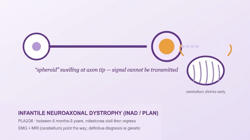
Infantile neuroaxonal dystrophy (INAD) is a progressive neurodegenerative disease beginning in infancy, caused by mutations in the PLA2G6 gene. The broad family named after this gene is called PLAN (PLA2G6-associated neurodegeneration): classic INAD lies at the severe end, atypical childhood-onset forms in the middle, and dystonia-parkinsonism (PARK14) at the adult end. The cellular hallmark of the disease is the formation of "spheroid" swellings at the tips of nerve fibres — these are points where axonal traffic is blocked and the signal cannot be transmitted. INAD is also one of the most common childhood members of the neurodegeneration with brain iron accumulation (NBIA) family; however, iron accumulation is a late finding, and its absence does not exclude the diagnosis.
Genetic Cause and Mechanism
- PLA2G6 encodes an enzyme (iPLA2β) that maintains the phospholipid balance of cell membranes. When function is lost, membranes cannot be repaired, and the mitochondria and axon terminals are damaged → axonal spheroids, cerebellar neuron loss and, over time, iron accumulation in the basal ganglia.
- Inheritance is autosomal recessive: both parents are carriers, and the recurrence risk in each pregnancy is 25% — genetic counselling and prenatal diagnosis options should be discussed with every family.
- The genotype-phenotype relationship is roughly as follows: variants that completely abolish enzyme function tend to cause classic INAD, while those leaving partial function tend to cause atypical/later-onset forms; variability can occur even within the same family.
Clinical Picture — Classic INAD
- Onset (6 months-3 years, usually around 1 year): In an infant whose development in the first year is mostly normal or mildly delayed, skills plateau and then regress — sitting deteriorates and new skills fail to appear. The typical early signature is marked truncal hypotonia.
- A "mixed" neurological signature: Because the disease affects both central and peripheral nerve terminals, the picture gradually turns into a combination of flaccidity and stiffness: deep tendon reflexes are lost (denervation) while the arms and legs become spastic, and the picture progresses towards symmetric tetraparesis/tetraplegia.
- Eye findings are early and point to the diagnosis: nystagmus, strabismus, and progressive optic atrophy (pale optic disc, reduced vision). Visual evoked potentials (VEP) become abnormal.
- Other features: In the later period, swallowing difficulty, loss of speech, cognitive impairment; in some cases (often at a late stage) seizures and fast rhythms on EEG; sleep disturbance and episodes of irritability (should be assessed together with a search for pain, reflux and constipation).
The Atypical Form and the PARK14 End
The atypical childhood form starts later (1.5-6 years) and progresses more slowly; ataxia, speech disorder and behavioural/autism-like features may predominate. At the adult end (PARK14), dystonia-parkinsonism plus psychiatric symptoms occur at a young age — the spectrum of the same gene should be kept in mind during family screening.
Diagnosis
- Two guiding investigations: EMG — findings of chronic denervation (anterior horn/axonal involvement; axonal pattern on nerve conduction) and brain MRI — early and marked cerebellar atrophy (+ signal change in the cerebellar cortex); over time, in some cases, iron accumulation in the globus pallidus (on SWI/T2*). Optic nerve thinning and abnormal VEP support the diagnosis.
- The definitive diagnosis is genetic: PLA2G6 sequence analysis/panel. Tissue biopsy (skin-nerve-conjunctiva) demonstrating axonal spheroids, once the former gold standard, has now been replaced by genetic testing.
- Pitfalls in differential diagnosis: (1) Onset with hypotonia plus areflexia may be mistaken for SMA — eye findings (nystagmus/optic atrophy) and cerebellar atrophy distinguish INAD; PLA2G6 should be considered in an SMN1-negative "SMA-like" infant. (2) Regression plus eye findings may be confused with NCL — in NCL, vision loss is of retinal origin (the ERG is extinguished) and seizures are an early, prominent feature; in INAD, optic atrophy plus EMG denervation is the signature (see the NCL guide). (3) Other NBIA members (PKAN, etc.) are distinguished by later age of onset and a different MRI pattern. For the general framework, see: distinguishing white from grey matter in the regressing child.
Treatment — An Honest Framework
- There is currently no approved treatment that stops the disease; the main treatment is multidisciplinary, anticipatory supportive care — and this care genuinely changes quality of life.
- Spasticity-dystonia: physiotherapy plus positioning-orthoses; baclofen, botulinum toxin when needed; screening for triggers (pain, reflux, constipation, urinary tract) in painful dystonic episodes.
- Nutrition-swallowing: consistency adjustment guided by swallowing assessments; gastrostomy should be discussed before the weight curve falls; reflux and constipation should be actively managed.
- Vision-communication: as vision declines, auditory-tactile communication and simple yes-no systems should be established early; if seizures develop, standard antiepileptics are used (there is no syndrome-specific contraindication).
- Respiratory: secretion management, early assessment in infection; decisions about respiratory support at an advanced stage should be discussed with the family in advance, together with the palliative care team.
- Research horizon: A deuterated fatty acid (RT001), aimed at making membrane lipids resistant to oxidative damage, has been trialled in INAD, but results have not shown definite benefit; gene therapy and enzyme-pathway approaches are at the preclinical stage. All of these are at the research stage; they are not a recommendation for clinical use — participation in patient registries (NBIA/INAD registries) is the route to future access to studies.
Follow-up — Summary
The backbone of follow-up: a developmental-skills map and updated goals every 3-6 months · nutrition-weight and swallowing assessment at every visit · annual eye assessment (optic atrophy/VEP) and a vision support plan · examination for spasticity-dystonia and hip-scoliosis (with radiography in the less mobile child) · screening for a source of pain in sleep disturbance and irritability · EEG if seizures develop · a respiratory-secretion plan · bone health (vitamin D; DEXA in case of fracture/immobility) · genetic counselling for every family (25%) and optional prenatal diagnosis · caregiver burden and early introduction to the palliative care team.
Prognosis — An Honest Framework for Families
- Classic INAD is progressive: skills are often lost markedly from 2-3 years of age onwards; many children reach a level requiring intensive care and support by school age, and life expectancy is often limited to the first decade — this is hard for a family to hear; it should be discussed early and with compassion, as it makes planning possible.
- In atypical forms, the course is markedly slower; it can extend into adolescence-adulthood.
- Goals are updated by stage: first preserving skills and communication, then comfort and freedom from pain, and a care routine the family can sustain. Well-established supportive care is the answer to the statement "there is much that can be done" in this diagnosis.
Daily Life
- Record skills and episodes on video; work with short-term goals of 3-6 months (sitting support, spoon use, pointing to choose).
- Keep the daily routine fixed; in episodes of irritability, first look for a source of pain (teeth, reflux, constipation, hip).
- Set up the positioning-orthosis routine at home with the physiotherapist; prevent pressure sores and pain.
- Discuss with your doctor whether to postpone genetic counselling when planning for siblings.
- NBIA/INAD family associations and registries: a resource for sharing experience and for research calls.